In the course of the follow-up visits (Nov 2011) to Wildlife (ANWC), Insect (ANIC) and Herbarium (ANH) curators and managers emphasise the way images and visualisation contribute to the development and management their collections. The Atlas of Living Australia, the remote microscope, microscopy, scanning, analysis techniques and DNA sequencing are central to expansion and revision of collections, the research and analysis of extant material as well as managing and accessing data attached to each specimen.
The significance of contemporary imaging technology for studying these collections and for revealing the relationship between them also underpins the concept of the CSIRO Spectra symposium and an exhibition proposed for later in 2012.
My visits continue to yield audio and visual material that highlights the scope and the intrinsic relationship between specimens and images, within and across the collections. Presented with so much extraordinary material in the course of these visits it maybe easy become overwhelmed —wondering at it all. To navigate this mass of specimens, images, and data I am tracing the ecology of a threatened Spider orchids and its symbiotic relationship to other specimens in the soil, plant, insect and wildlife collections. Unraveling its association with other organisms such as the mycorrhizal fungi and identifying its pollinator is not as straightforward as it may first appear. The ecology of this and other orchids is elaborate and their capacity for sophisticated deception of particular pollinators is an intriguingly complex feature of their ecology.
Identification and analysis of different aspects of the orchid’s ecology reflect the scope of visualising technologies. Spatial and spectral information about the biochemistry of different organic material, such as pheromones or soil types, presents in very different visual systems from (numerical data in) graphs to satellite maps and these converge in a whole account of this specimen.
While the development of botany and other sciences can be linked (historically) to the use of visually accurate images and universally accepted graphic conventions: for artists and scientists digital tools and techniques of visualisation offer very different possibilities for observation and experience. They not only reveal a paradoxical relationship between visual accuracy and truth, they radically change the way that images are regarded in both specialist areas and in the broader culture.
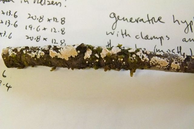
At the Australian National Herbarium’s Cryptogam collection (ferns, mosses, liverworts and lichens, algae and fungi) housed at the Australian National Botanical Gardens (ANBG), I see how the small and often unspectacular dried specimens are handled and organised. Heino Lepp, whose special study is mycology (fungi), talks about his research and the co-existence of analogue and digital processes —drawing writing and DNA analysis are used to describe and identify.
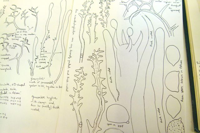
Heino’s drawings are detailed descriptions of specimens through close observation of their form and structure. To see and draw the microscopic detail of the corticioid fungi Heino uses a camera lucida, a long-standing aid in scientific illustration. The black line drawings of this specimen show it fragmented and these irregular forms and structures are drawn at much larger scale than the specimen.
Corticioid fungi forms on the underside of wood and in a comparison of drawings in different accounts from America, Europe, Asia and New Zealand, it is interesting to note first that these specimens are drawn using detailed rendering techniques that gives them a dense, rigid almost architectural structure and the respective accounts are alternatively orientated growing up (from the wood), and in others the in others the wood is above and the fungi is hanging downs below it.
By contrast Heinio’s almost whimsical cartoon like drawings some with curly, rounded, pointed features show as much irregularity and variation in the specimen as it is possible to observe. The magnified shapes float informally across the drawing interspersed with hand written annotations describing different features. These drawn and written descriptions distinguish the specimen but it is clear how significant DNA is in further analysis and identification when species share similar features.
At the insect Collection (ANIC) Nicole Fisher who manages the imaging of the insect collection explains the significance of microscope techniques that enable automatic analysis and recognition of pollen grains taken from bees in the cabinets. In this instance the plant material found on bees in the collection can enable an isolated specimen to be reconnected with information about its environmental history and ecology. While this may tease the boundary between systematic separation of organic material into distinct collections, the high resolution microscopy images of the parasitic wasps (October) and scanning of whole draws (trays) of specimens, are among the numerous technologies that are re-imaging these collections.